Throughout even essentially the most routine visits, physicians hearken to sounds inside their sufferers’ our bodies — air shifting out and in of the lungs, coronary heart beats and even digested meals progressing by means of the lengthy gastrointestinal tract. These sounds present useful details about an individual’s well being. And when these sounds subtly change or downright cease, it might probably sign a major problem that warrants time-sensitive intervention.
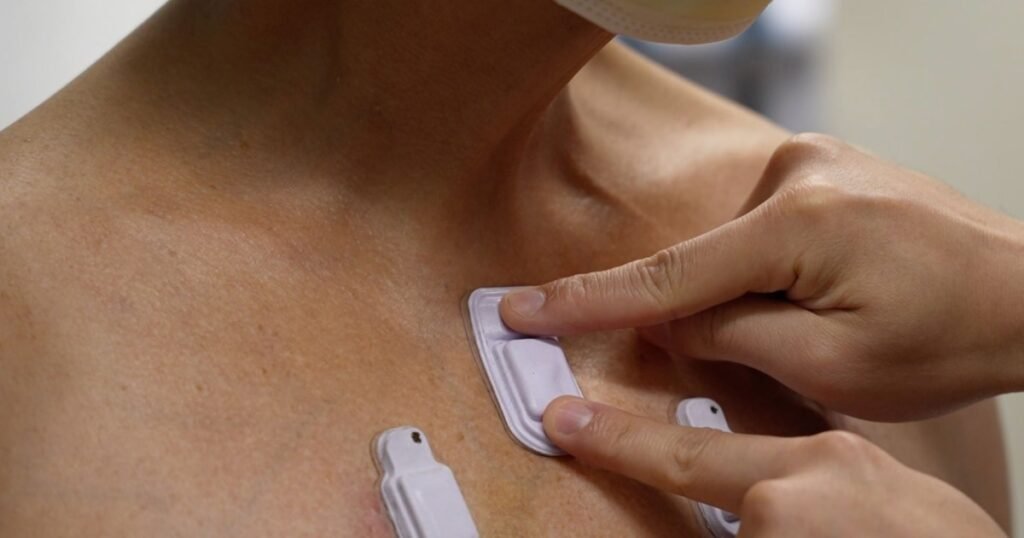
Now, Northwestern College researchers are introducing new comfortable, miniaturized wearable gadgets that go effectively past episodic measurements obtained throughout occasional physician exams. Softly adhered to the pores and skin, the gadgets repeatedly monitor these delicate sounds concurrently and wirelessly at a number of places throughout almost any area of the physique.
The brand new research was printed at this time (Nov. 16) within the journal Nature Medication.
In pilot research, researchers examined the gadgets on 15 untimely infants with respiratory and intestinal motility problems and 55 adults, together with 20 with continual lung illnesses. Not solely did the gadgets carry out with clinical-grade accuracy, in addition they supplied new functionalities that haven’t been developed nor launched into analysis or medical care.
“At the moment, there aren’t any present strategies for repeatedly monitoring and spatially mapping physique sounds at dwelling or in hospital settings,” mentioned Northwestern’s John A. Rogers, a bioelectronics pioneer who led the system improvement. “Physicians should put a traditional, or a digital, stethoscope on completely different components of the chest and again to hearken to the lungs in a point-by-point trend. In shut collaborations with our medical groups, we got down to develop a brand new technique for monitoring sufferers in real-time on a steady foundation and with out encumbrances related to inflexible, wired, cumbersome expertise.”
“The thought behind these gadgets is to offer extremely correct, steady analysis of affected person well being after which make medical selections within the clinics or when sufferers are admitted to the hospital or connected to ventilators,”mentioned Dr. Ankit Bharat, a thoracic surgeon at Northwestern Medication, who led the medical analysis within the grownup topics. “A key benefit of this system is to have the ability to concurrently pay attention and examine completely different areas of the lungs. Merely put, it’s like as much as 13 extremely skilled medical doctors listening to completely different areas of the lungs concurrently with their stethoscopes, and their minds are synced to create a steady and a dynamic evaluation of the lung well being that’s translated right into a film on a real-life laptop display.”
Rogers is the Louis Simpson and Kimberly Querrey Professor of Supplies Science and Engineering, Biomedical Engineering and Neurological Surgical procedure at Northwestern’s McCormick Faculty of Engineering and Northwestern College Feinberg Faculty of Medication. He additionally directs the Querrey Simpson Institute for Bioelectronics. Bharat is the chief of thoracic surgical procedure and the Harold L. and Margaret N. Technique Professor of Surgical procedure at Feinberg. Because the director of the Northwestern Medication Canning Thoracic Institute, Bharat carried out the primary double-lung transplants on COVID-19 sufferers within the U.S. and began a first-of-its-kind lung transplant program for sure sufferers with stage 4 lung cancers.
Complete, non-invasive sensing community
Containing pairs of high-performance, digital microphones and accelerometers, the small, light-weight gadgets gently adhere to the pores and skin to create a complete non-invasive sensing community. By concurrently capturing sounds and correlating these sounds to physique processes, the gadgets spatially map how air flows into, by means of and out of the lungs in addition to how cardiac rhythm adjustments in assorted resting and energetic states, and the way meals, fuel and fluids transfer by means of the intestines.
Encapsulated in comfortable silicone, every system measures 40 millimeters lengthy, 20 millimeters large and eight millimeters thick. Inside that small footprint, the system incorporates a flash reminiscence drive, tiny battery, digital elements, Bluetooth capabilities and two tiny microphones — one dealing with inward towards the physique and one other dealing with outward towards the outside. By capturing sounds in each instructions, an algorithm can separate exterior (ambient or neighboring organ) sounds and inside physique sounds.
“Lungs do not produce sufficient sound for a traditional particular person to listen to,” Bharat mentioned. “They only aren’t loud sufficient, and hospitals will be noisy locations. When there are folks speaking close by or machines beeping, it may be extremely troublesome. An necessary side of our expertise is that it might probably appropriate for these ambient sounds.”
Not solely does capturing ambient noise allow noise canceling, it additionally gives contextual details about the sufferers’ surrounding environments, which is especially necessary when treating untimely infants.
“No matter system location, the continual recording of the sound atmosphere gives goal knowledge on the noise ranges to which infants are uncovered,” mentioned Dr. Wissam Shalish, a neonatologist on the Montreal Youngsters’s Hospital and co-first writer of the paper. “It additionally affords fast alternatives to handle any sources of traumatic or probably compromising auditory stimuli.”
Non-obtrusively monitoring infants’ respiratory
When creating the brand new gadgets, the researchers had two weak communities in thoughts: untimely infants within the neonatal intensive care unit (NICU) and post-surgery adults. Within the third trimester throughout being pregnant, infants’ respiratory techniques mature so infants can breathe exterior the womb. Infants born both earlier than or within the earliest levels of the third trimester, due to this fact, usually tend to develop lung points and disordered respiratory problems.
Significantly frequent in untimely infants, apneas are a number one explanation for extended hospitalization and probably dying. When apneas happen, infants both don’t take a breath (attributable to immature respiratory facilities within the mind) or have an obstruction of their airway that restricts airflow. Some infants may also have a mixture of the 2. But, there aren’t any present strategies to repeatedly monitor airflow on the bedside and to precisely distinguish apnea subtypes, particularly in these most weak infants within the medical NICU
